Zooming


Zooming |
|
|
A number of ways of zooming are available in Mnova.
The most common type of zooming in spectroscopy applications is the 'Zoom In' mode. This mode can be entered by following the menu 'View/Zoom/Zoom In' or by using the 'Zoom In' icon on the toolbar.
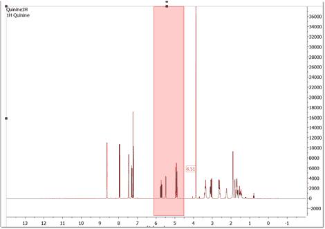
In this area, Mnova presents a significant innovation, allowing the user to apply 'horizontal', 'vertical' and 'box' zooms, as illustrated by the figures below. Horizontal Zoom. Click the 'Zoom In' icon on the toolbar (or press <Z> key), hold down the left mouse button on the spectrum display and drag the magnifying glass cursor (with a red horizontal segment) across the area you want to zoom in to; a highlighted rectangle indicating the new spectral region to be displayed once the mouse is released, will follow the cursor. Use 'Undo' to revert to the previous display. A zoom lens of a 20% will be obtained by clicking on the left mouse button.
The zoom cursor shows information about the position (same units as in spectrum scales). Once the starting position is selected, only the values for the ending position (f1, f2 or both dimensions, depending on the type of the zoom applied) are displayed.
Please note that holding down 'Ctrl+Space bar' (in Mac: 'Space+Cmd') will activate the zoom in temporarily.
A fixed zoom area can be moved around the spectra using Alt+Arrow keys
Vertical Zoom. Click the 'Zoom In' icon on the toolbar and then press the <Z> key (or just press twice the 'Z' key). You will see a red vertical line segment beside the mouse pointer) while the cursor is located on the spectrum display and then click and drag the cursor vertically over the area you want to zoom in on; once again you will see highlighted red rectangle.
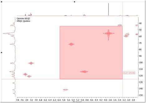
Box Zoom. Click the 'Zoom In' icon on the toolbar and then press <Z> key twice with the mouse pointer on the spectrum display (or just press three times the 'Z' key), then click and drag the cursor diagonally over the area you want to zoom in (highlighted rectangle in red). This zoom is very useful for 2D-NMR.
In order to switch between these three types of zoom, just press the <Z> key whilst in the 'Zoom In' mode.
Zoom Out: Click the 'Zoom Out' icon on the toolbar and click on the left mouse button to obtain a zoom lens out of a 20%.
Back/Forward Zoom: You can back/forward to previous/next zoom(s) level(s) by using the shortcuts: Shift+LeftArrow(previous)/Shift+RightArrow(next) or the buttons "Previous/Next Zoom".
Manual Zoom: Click 'Manual Zoom' on the toolbar or by following the menu 'View/Zoom/Manual Zoom'. The manual zoom is slightly different in 1D to that in 2D NMR spectra where you may select spectral spectral limits for both dimensions.
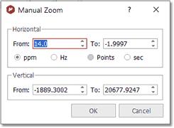
1. If you are working on a 1D-NMR spectrum, a dialog box like this will open:
You can select the limits of the spectral region of interest in ppm, Hz, points or sec and also for the intensity.
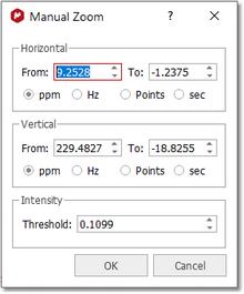
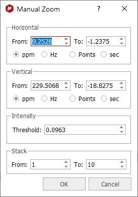
2. If you are working on a 2D-NMR spectrum, the dialog box will look like this:
Select the horizontal, vertical limits and intensity of the region of the spectrum that you want to zoom in on, and click 'OK'.
In stacked plots, you will be also able to select the traces to be used:
OTHER ZOOMING MODES 1. FULL SPECTRUM: This tool may be used to revert to a full spectrum from a zoomed view. This may be done from the menu 'View/Zoom/Full Spectrum' or by using the 'Full Spectrum' icon
2. FIT TO HIGH: The Fit to High function adjusts the display of spectral intensity in both 1D and 2D spectra so that the strongest signal is optimally displayed.
3. FIT TO HIGHEST COMPOUND: The Fit to Highest function adjusts the display of spectral intensity in both 1D and 2D spectra so that the strongest compound signal is optimally displayed (discarding solvent or impurity peaks). You can select this mode by default from the 'File/Preferences/NMR/Import' menu. 4. PANNING: While the spectrum is in a zoomed state, click on the 'PAN' icon
You will be able to toggle the Full View window by using the F2 key. This will allow you to get a 'Super Fast' panning by using the Full View window.
You could also use the 'Alt' key plus arrow keys (up&down) to apply a fixed panning in 2D-NMR.
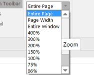
4. ZOOMING COMBO BOX: In addition to the traditional 'Zoom In' mode, Mnova also allows the user to apply different zooms, including Zoom Out (from a zoomed state), by using the zooming combo box.
These zooming modes are:
✓Percentage zooms: These allow different size views of the object, by selecting the different percentages available (from 400 to 25%). ✓Entire Page: This is the standard view, which can be seen in all the figures above. ✓Page Width: In this view, the program will fit the size of the paper to the window width, whilst preserving the original paper proportions. ✓Entire Window: This is a special view within Mnova. It will fit the size of the paper to the size of the available window, to optimize the available screen area. This view represents an exception to the WYSIWYG concept, since in this case the proportions of the screen paper may differ from the proportions of the printer paper and therefore of what you will get when printing the page.
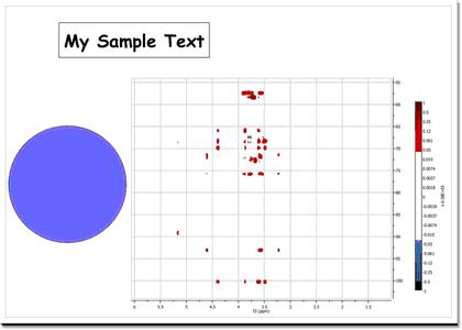
The Entire Window concept deserves further comments. For example, consider the following scenario in which the page zooming mode is set to Entire Page:
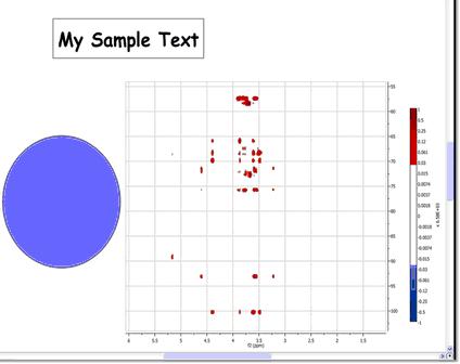
As seen in the figure above, Mnova is a fully WYSIWYG application. This can be appreciated in the blue circle which was created with the ellipse tool and then its dimensions set to form an actual circle. However, in this case there is a significant space of the window which is not covered by the page (see gray regions surrounding the page) which can be considered as wasted space. In order to make the most of the available space, we can use the so-called Entire Window zooming mode. In this mode, the page stretches to fit all the available space. Of course, this means that paper proportions (i.e., width/height ratio) have to be changed and thus the WYSIWYG principle is broken. As always, a picture is worth a thousand words:
Note that all the objects are now distorted. However, this mode is very convenient to work with spectra on the screen as the graphical spectral proportions are rarely an issue. Nevertheless, if you print a document in this non WYSIWYG mode, you will get exactly the same results as when working on real WYSIWYG modes.
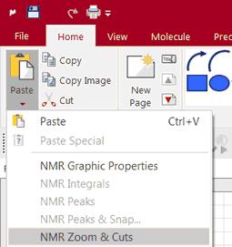
Please bear in mind that you can copy the zoom areas from one spectrum to another just by doing Ctrl/Cmd+C in the first one and 'Home/Paste/NMR Zoom&Cuts':
|